Dental professionals utilize radiographs to detect, diagnose, treat, and monitor oral conditions and diseases.
Digital radiography in Dentistry is a form of imaging using digital sensors in the mouth. Digital imagery enhances teeth, gums, and other structures and conditions in a patient’s mouth.
Women: Always tell your Doctor if there is a possibility that you are pregnant and if you are currently breast feeding. Removing jewelry, eye glasses and metal objects is important as they might interfere with the x-ray imaging.
Panoramic (PAN)

A panoramic x-ray uses a small dose of ionizing radiation. It is commonly performed by Dentists and Oral Surgeons in everyday practice. Panos may be used with a Treatment Plan for dentures, braces, extractions and implants. Malcomson Dentistry has a two-dimensional (2-D) PAN that captures the entire mouth in a single image, including the teeth, upper and lower jaws, surrounding structures and tissues.
The jaw is a curved structure similar to that of a horseshoe. The panoramic x-ray produces a flat image of a curved structure. An x-ray is a non-invasive medical procedure, that helps physicians diagnose and treat medical conditions. X-rays are the oldest and most frequently used form of medical imaging. Unlike a traditional intraoral x-ray where the film/x-ray detector is placed inside of the mouth, the digital photo for a panoramic x-ray is in the machine.
What are some common uses of a Panoramic x-ray?
Covering a wider area than a conventional intraoral x-ray resulting in valuable information about the maxillary sinuses, tooth positioning and other bone abnormalities. A panoramic x-ray can reveal dental and medical problems such as: advanced periodontal disease, cysts in the jaw bones, jaw tumors and oral cancer, impacted teeth including wisdom teeth, jaw disorders (also known as temporomandibular joint or TMJ disorders) and sinusitis.
Full Mouth Series (FMX)
A “full-set” of periapical x-rays show all of your teeth, surrounding bone and help to diagnose cavities, cysts or tumors, abscesses, impacted teeth and gum disease. Consisting of 14-20 individual x-rays, a FMX is typically recommended during one of your first visits with a new Dentist to aid in proper diagnosis and treatment planning.

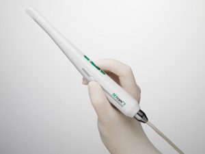
Digital Intra-Oral Camera and Cari-Vu Transilluminator
The ability to see subtleties using digital radiography is crucial to diagnosis, collaboration, and patient communication. Using an Intra-Oral Camera and Transilluminator combine advanced technologies to deliver clinically meaningful images that are extremely clear and highly detailed. Fancy words, but a simple concept: patients benefit from a reduction in the number of exposure-related dental radiograph retakes.
When a suspicious area is identified on a radiograph, a transilluminated image can reveal the extent of the damage and help to confidently determine treatment. Transillumination can show lesions in the beginning stages, using CariVu during routine prophylaxis can help to identify questionable areas early on and decide on a course of preventive care. When used together; a radiograph, a transilluminated image and an intra-oral photo can provide a comprehensive picture of the health of a tooth. When a suspicious area is identified on a radiograph, a transilluminated image can reveal the extent of the damage and help to confidently determine treatment. Transillumination can show the beginning stages of lesions.

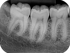
Bitewing Radiographs (BWX)
Bitewings (BWX) are the most common form of dental x-rays. BWX show the upper and
lower teeth above the gumline and the height of the bone between the teeth. They can help
diagnose gum disease and cavities between teeth. Typically, BWX are taken once a year
and are highly recommended for patients who have frequent decay and less often for those
with little decay. Dr. Malcomson evaluates the necessity on a patient basis.
Periapical (PA)